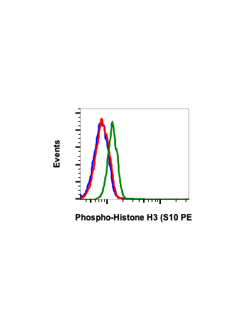
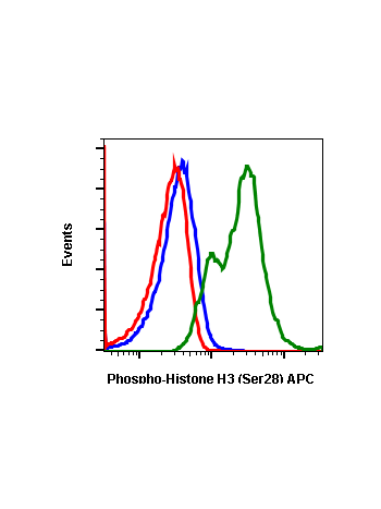
Phospho-Histone H3 (Ser10) (4B6) rabbit mAb APC conjugate
From
$118.80
In stock
Only %1 left
SKU
2064
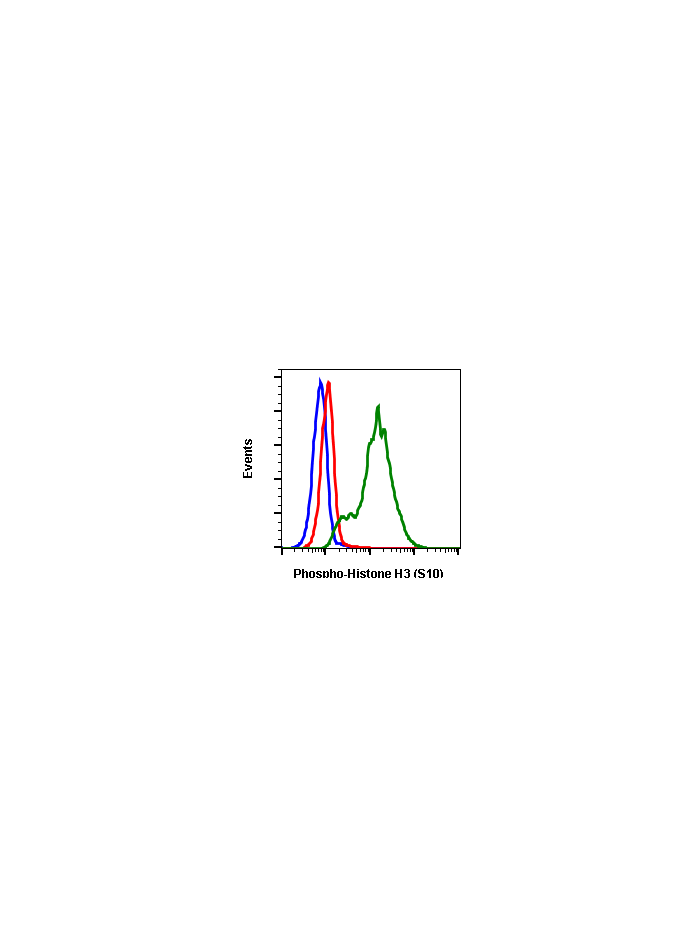
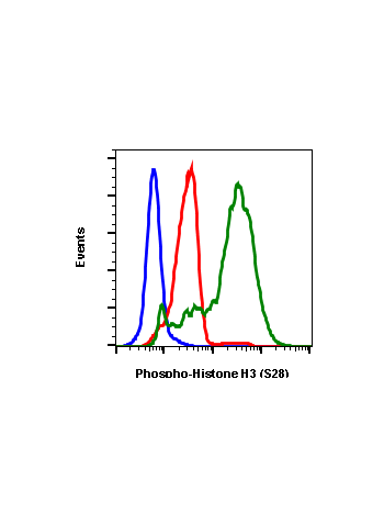
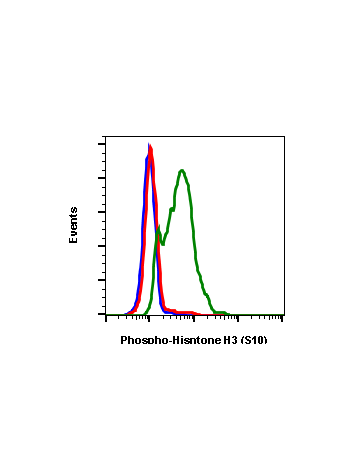
Histones are highly conserved proteins that serve the core of nucleosomes, which serve to organize chromatin fiber for DNA packing. Histone H3 phosphorylation plays a major role in both transcriptional activation, which requires unpacking of the chromatin structure, and in chromosome packing during cell division. Histone H3 is phosphorylated at residues Ser10 and Ser28, and is acetylated at Lys14. Phosphorylation at Ser10 occurs during entry into mitosis prior to chromatin condensation, and phosphorylation at Ser28 follows a similar pattern. In response to EGF stimulation, it has been proposed that sequential Ser10 phosphorylation, then Lys14 acetylation occurs, causing a change in chromatin structure and gene activation.
| Applications | Flow Cytometry |
|---|---|
| Clone | HisH3S10-4B6 |
| Format | APC |
| Validated Reactivity | Human |
| Cross Reactivity | Predicted to work with mouse, rat and other homologues. |
| Clonality | Monoclonal |
| Immunogen | A synthetic phospho-peptide corresponding to residues surrounding Ser10 of human phospho histone H3 |
| Formulation | 1X PBS, 0.09% NaN3, 0.2% BSA |
| Isotype | Rabbit IgGk |
| Preparation | Protein A+G |
| Recommended Usage | For flow cytometric staining, the suggested use of this reagent is 5 µL per million cells or 5 µL per 100 µL of staining volume. It is recommended that the reagent be titrated for optimal performance for each application. |
| Storage | 2-8ºC |
| Pseudonyms | Histone H3.1t, H3t, H3FT, HIST3H3 |
| Uniprot ID | Q16695 |
| References | Hans F and Dimitrov S. (2001) Oncogene. 20: 3021-3027. Cheung P, Tanner KG, Cheung WL, et al. (2000) Molecular Cell. 5: 905-915. |
Write Your Own Review