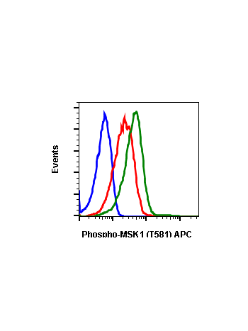
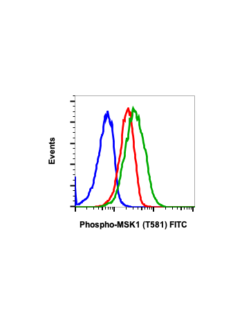
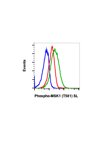
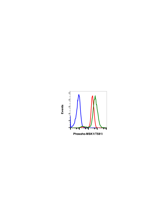
Phospho-MSK1 (Thr581) (A5) rabbit mAb
Product Overview
Description
MSK1 (mitogen and stress activated protein kinase 1, phospho MSK1) is activated by Erk in response to growth factors and by and p38 in response to cellular stress (1). MSK1 is similar to RSK1 in that it has two kinase domains and a connecting regulatory linker region (2). S364/S381 phosphorylation activates RSK1 (3), which is analogous to residues S360 and S376 of MSK1, which may be important for phospho MSK1 activity.
Product Details
Specification | Description |
---|---|
Applications | Flow Cytometry, WB |
Clone | MSK1T581-A5 |
Format | Unconjugated |
Validated Reactivity | Human, Mouse, Rat |
Cross Reactivity | Predicted to work with mouse, rat and other homologues. |
Detection | Anti-Rabbit IgG |
Clonality | Monoclonal |
Immunogen | A synthetic phospho-peptide corresponding to residues surrounding Thr581 of human phospho MSK1 |
Formulation | 1X PBS, 0.02% NaN3, 50% Glycerol, 0.1% BSA |
Isotype | Rabbit IgGk |
Preparation | Protein A+G |
Recommended Usage | 1µg/mL – 0.001µg/mL. It is recommended that the reagent be titrated for optimal performance for each application. See product image legends for additional information. |
Storage | -20ºC |
Pseudonyms | Ribosomal protein S6 kinase alpha-5, S6K-alpha-5, 90 kDa ribosomal protein S6 kinase 5, Nuclear mitogen- and stress-activated protein kinase 1, RSK-like protein kinase, RSKL, RPS6KA5 |
Uniprot ID | O75582 |
References | 1. Deak, M. et al. (1998) EMBO J. 17, 4426-4441. 2. Pierrat, B. et al. (1998) J. Biol. Chem. 273, 29661-29671. 3. Dalby, K.N. et al. (1998) J Biol Chem 273, 1496-505. 4. Markou, T. and Lazou, A. (2002) Biochem J 365, 757-63. |
Images

Peptide blocking flow cytometric analysis of C6 cells secondary antibody only negative control (light blue) or pervanadate-treated and stained using 0.1µg/mL isotype control Cat. #2141 (orange) or pervanadate-treated and stained using 0.1µg/mL Phospho-MSK1 (Thr581) antibody MSK1T581-A5 Cat. #2181 (green) or pervanadate and blocked with phospho peptide (gold) or pervanadate and blocked with non-phospho peptide (purple).

Flow cytometric analysis of C2C12 cells secondary antibody only negative control (blue) or 0.1 µg/mL of isotype control Cat. #2141 (orange) or treated with imatinib (red) or with pervanadate (green) using Phospho-MSK1 (Thr581) antibody MSK1T581-A5 at 0.1 µg/mL. Cat #2181.

Peptide blocking flow cytometric analysis of K562 cells secondary antibody only negative control (light blue) or imatinib-treated (red) or pervanadate-treated (green) or imatinib and blocked with phospho-peptide (black) or pervanadate and blocked with phospho peptide (gold) or imatinib and blocked with non-phospho peptide (dark blue) or pervanadate and blocked with non-phospho peptide (purple) Phospho-MSK1 (Thr581) antibody MSK1T581-A5 at 0.1µg/mL. Cat. #2181.

Western blot analysis of 293T cell extract untreated or treated with UV using 0.1 ug/mL Phospho-MSK1 (Thr581) antibody MSK1T581-A5. Cat. #2181.

Flow cytometric analysis of U937 cells secondary antibody only negative control (blue) or untreated (red) or treated with CalA (green) using Phospho-MSK1(T581) antibody MSK1T581-A5 1.0 µg/mL. Cat. #2181.

Western blot analysis of 293T cell extract untreated or treated with staurosporine using 0.1 µg/mL Phospho-MSK1 (Thr581) antibody MSK1T581-A5 Cat. #2181 or Company C antibody at 0.02 µg/mL (manufacturer’s recommended concentration) developed using the same exposure.