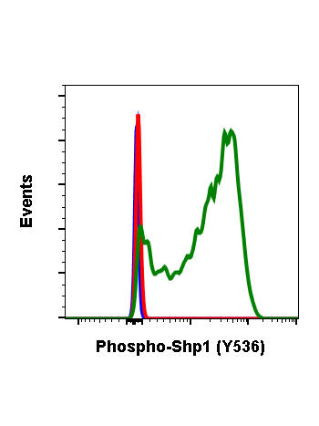
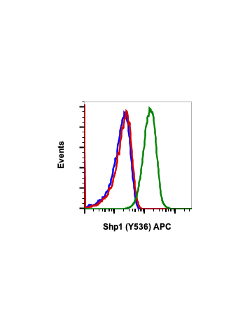
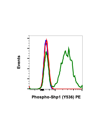
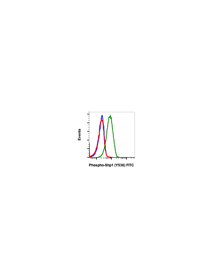
Phospho-Shp1 (Tyr536) (2A7) rabbit mAb FITC conjugate
Src homology-2 domain containing phosphatase-1 (Shp1) typically functions as a negative regulator for signal transduction, interacting with Zap70, CD5, CD3ε, and IL-2R in T cells. Shp1 suppresses cellular signaling by dephosphorylating target proteins. Shp1 is expressed predominantly in hematopoietic cells, where it can plays a role in malignant transformation into cancer cells. Methylation of the Shp1 promoter has been shown to silence Shp1 expression and promote the development of cancer. Shp1 negatively regulates PDGF and insulin tyrosine receptor pathways, and negatively regulates Lck in the TCR signaling pathway to limit TCR responsiveness. Shp1 activity is prevented by TAOK3 in T cells to allow for T cell activation and differentiation.
| Applications | Flow Cytometry |
|---|---|
| Clone | Shp1Y536-2A7 |
| Format | FITC |
| Validated Reactivity | Human |
| Cross Reactivity | Predicted to work with mouse, rat and other homologues. |
| Clonality | Monoclonal |
| Immunogen | A synthetic phospho-peptide corresponding to residues surrounding Tyr536 of human phospho Shp1 |
| Formulation | 1X PBS, 0.09% NaN3, 0.2% BSA |
| Isotype | Rabbit IgGk |
| Preparation | Protein A+G |
| Recommended Usage | For flow cytometric staining, the suggested use of this reagent is 5 µL per million cells or 5 µL per 100 µL of staining volume. It is recommended that the reagent be titrated for optimal performance for each application. See product image legends for additional information. |
| Storage | 2-8ºC |
| Pseudonyms | Tyrosine-protein phosphatase non-receptor type 6, Hematopoietic cell protein-tyrosine phosphatase, Protein-tyrosine phosphatase 1C, PTP-1C, SH-PTP1, PTPN6, HCP, PTP1C |
| Uniprot ID | P29350 |
| References | 1. Oka T, Ouchida M, Koyama M, et al. (2002) Cancer Research. 62:6390-6394. 2. Geraldes P, Hiraoka-Yamamoto J, Matsumoto M, et al., (2009) Nature Medicine. 15:1298-1306. 3. Ormonde JVS, Li Z, Stegen C, and Madrenas J. (2018) Journal of Immunology 201:3431-3442. |