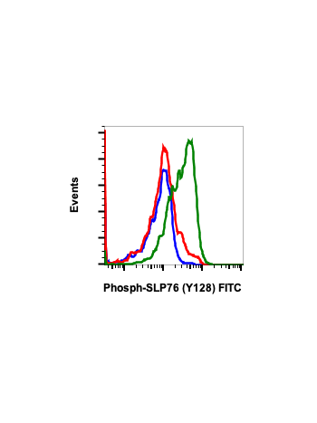
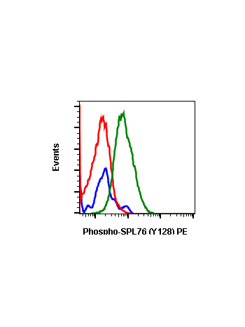
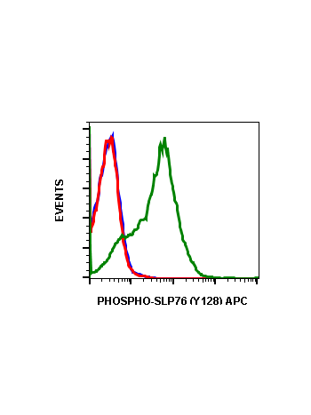
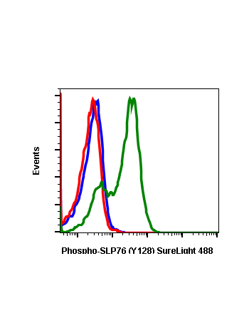
Phospho-SLP-76 (Tyr128) (3F8) rabbit mAb
From
$210.00
In stock
Only %1 left
SKU
2136
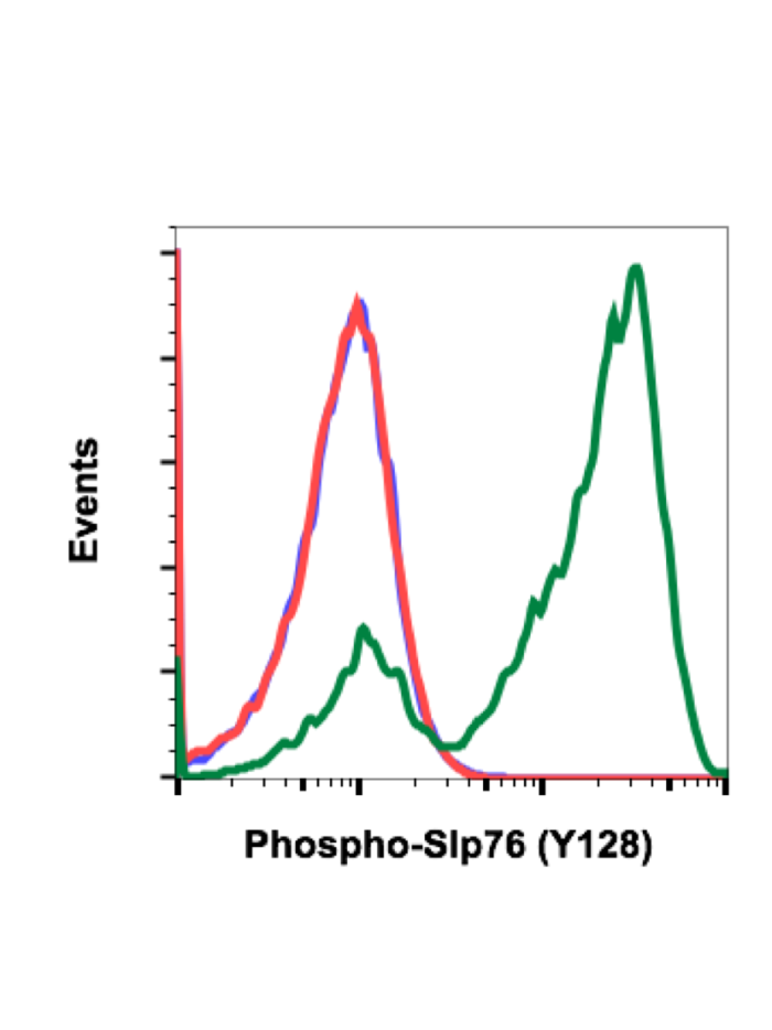
SH2 Domain-Containing Leukocyte Protein Of 76 KDa (SLP-76) is an adaptor protein that plays a role in signal transduction in T cells. Studies using a SLP-76-deficient T cell line have demonstrated that SLP-76 is required for optimal phosphorylation and activation of both PLCγ1 and the Ras pathway. SLP-76 phosphorylation is mediated by Zap70 upon TCR stimulation. Within an N-terminal acidic region, SLP-76 possesses three tyrosines (Tyr113, 128, and 145), which are phosphorylated upon activation. The sterile α-motif (SAM) domain of SLP-76 drives formation of dimers and higher order oligomers. SLP-76 micro-clusters at the immunological synapse enhance signal transduction and T cell activation.
| Applications | Flow Cytometry |
|---|---|
| Clone | SLP76Y128-3F8 |
| Format | Unconjugated |
| Validated Reactivity | Human, Mouse |
| Cross Reactivity | Predicted to work with mouse, rat and other homologues. |
| Detection | Anti-Rabbit IgG |
| Clonality | Monoclonal |
| Immunogen | A synthetic phospho-peptide corresponding to residues surrounding Tyr128 of human phospho SLP-76 |
| Formulation | 1X PBS, 0.02% NaN3, 50% Glycerol, 0.1% BSA |
| Isotype | Rabbit IgGk |
| Preparation | Protein A+G |
| Recommended Usage | 1µg/mL – 0.001µg/mL. It is recommended that the reagent be titrated for optimal performance for each application. See product image legends for additional information. |
| Storage | -20ºC |
| Pseudonyms | Lymphocyte cytosolic protein 2, SH2 domain-containing leukocyte protein of 76 kDa, SLP76, LCP2 |
| Uniprot ID | Q13094 |
| References | Zhang MS, Tran PM, Wolff AJ, Tremblay MM, Fosdick MG, and Houtman JCD. (2018) Science Signaling. 11:eaam9095. Yablonski D, Kuhne MR, Kadlecek T, and Weiss A. (1998) Science. 281:413-416. Thaker YR, Recino A, Raab M, Jabeen A, Wallberg M, Fernandez N, and Rudd CE. (2017) Journal of Biological Chemistry. 292:6281-6290. |
Write Your Own Review