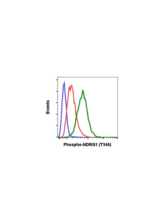
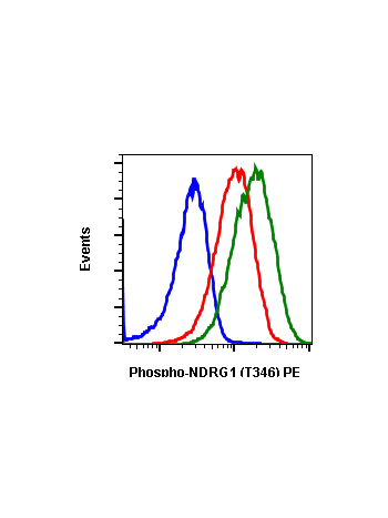
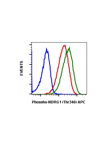
Phospho-NDRG1 (Thr346) (F5) rabbit mAb
From
$210.00
In stock
Only %1 left
SKU
2106
N-Myc down-regulated gene 1 (NDRG1) has been reported to be a direct transcriptional target of p53. NDRG1 appears to play a necessary, but not sufficient, role in apoptosis, though its exact mechanism of action remains unknown. NDRG1 expression is elevated in non-small cell lung cancer cells, promoting cancer growth and reducing cytotoxicity to certain anti-cancer drugs. NDRG1 is also elevated in solid tumors and is recognized as a negative prognostic indicator in breast cancer. Elevated NDRG1 expression is correlated with disease recurrence and metastasis in breast cancer. NDRG1 is phosphorylated by Sgk1, which itself is activated by mTORC2. Phosphorylation of NDRG1 at Thr346 promotes cellular differentiation in adipocytes.
| Applications | Flow Cytometry, WB |
|---|---|
| Clone | NDRG1T346-F5 |
| Format | Unconjugated |
| Validated Reactivity | Human, Mouse |
| Cross Reactivity | Predicted to work with mouse, rat and other homologues. |
| Detection | Anti-Rabbit IgG |
| Clonality | Monoclonal |
| Immunogen | A synthetic phospho-peptide corresponding to residues surrounding Thr346 of human phospho NDRG1 |
| Formulation | 1X PBS, 0.02% NaN3, 50% Glycerol, 0.1% BSA |
| Isotype | Rabbit IgGk |
| Preparation | Protein A+G |
| Recommended Usage | 1µg/mL – 0.001µg/mL. It is recommended that the reagent be titrated for optimal performance for each application. See product image legends for additional information. |
| Storage | -20ºC |
| Pseudonyms | Differentiation-related gene 1 protein, N-myc downstream-regulated gene 1 protein, Nickel-specific induction protein Cap43, Reducing agents and tunicamycin-responsive protein, RTP, Rit42 |
| Uniprot ID | Q92597 |
| References | Stein S, Thomas EK, Herzog B, Westfall MD, Rocheleau JV, Jackson II RS, Wang M, and Liang P. (2004) Journal of Biological Chemistry. 279:48930-48940. Du A, Jiang Y, and Fan C. (2018) International Journal of Medical Sciences. 15:1502-1507. Cai K, El-Merahbi R, Loeffler M, Mayer AE, and Sumara G. (2017) Scientific Reports 7:7191. Sevinsky CJ, Khan F, Kokabee L, Darehshouri A, Maddipati KR, and Conklin DS. (2018) Breast Cancer Research. 20:55. |
Write Your Own Review


