Phospho-Histone H2A.X (Ser139) (1E4) rabbit mAb

Catalog #: 2231
Select Your Size Price
200 uL (0.5 mg/mL)(2231-200) $570.00
20 uL (0.5 mg/mL)(2231-20) $210.00
From $210.00

Need to make a custom order?

Contact us

Note

20 μL is enough antibody for at least 20 Western blots.

Product Overview

Description
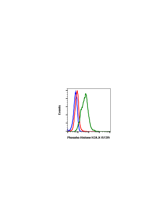
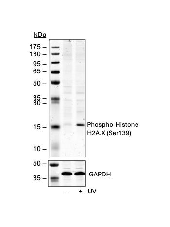
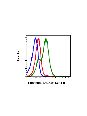
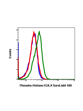
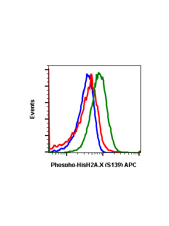
Histone H2AX is a variant of the nucleosome core histone H2A and is phosphorylated at Ser139 in response to DNA damage. Histone H2AX phosphorylation is considered a specific reporter of double-strand DNA breaks. The protein is also referred to as γH2AX when phosphorylated at Ser139. H2AX phosphorylation is especially strong in response to double-strand breaks formed during apoptosis. However, physiological phosphorylation of Histone H2AX occurs when double-strand DNA breaks are formed during meiosis and V(D)J recombination. A549 and DU145 cell lines have been found to have higher expression levels of phosphorylated Histone H2AX compared to Jurkat, MCF-7, or HL-60 cell lines.

Product Details

Product Details
Specification Description
Applications Flow Cytometry, WB
Clone HisH2AXS139-1E4
Format Unconjugated
Validated Reactivity Human, Mouse
Cross Reactivity Predicted to work with mouse, rat, and other homologues.
Detection Anti-Rabbit IgG
Clonality Monoclonal
Immunogen A synthetic phospho-peptide corresponding to residues surrounding Ser139 of human phospho histone H2A.X.
Formulation 1X PBS, 0.02% NaN3, 50% Glycerol, 0.1% BSA
Isotype Rabbit IgGk
Preparation Protein A+G
Recommended Usage 1µg/mL – 0.001µg/mL. It is recommended that the reagent be titrated for optimal performance for each application. See product image legends for additional information.
Storage -20ºC
Pseudonyms H2AFX, H2AX, γH2AX, gamma-H2AX
Uniprot ID P16104
References Tanaka T, Halicka D, Huang X, et al. (2006) Cell Cycle. 5: 1940-1945.

Images

Cell Culture Products
Flow cytometric analysis of 3T3 cells secondary antibody only (blue) or untreated with 0.1 µg/mL of isotype control Cat. #2141 (orange) or untreated (red) or UV and PMA-treated (green) using phospho-Histone H2A.X (Ser139) antibody HisH2AXS139-1E4 Cat. #2231.
Cell Culture Products
Peptide blocking flow cytometric analysis of 293T cells secondary antibody only negative control (blue) or untreated (red) or treated with UV and PMA (green) or untreated and blocked with phospho-peptide (black) or treated and blocked with phospho peptide (gold) or untreated and blocked with non-phospho peptide (dark blue) or treated and blocked with non-phospho peptide (purple) using Phospho-Histone H2A.X (Ser139) antibody HisH2AXS139-1E4 at 0.1 µg/mL. Cat. #2231.
Cell Culture Products
Western blot analysis of HEK293T cell extract untreated or treated with UV using 0.05 µg/mL Phospho-Histone H2A.X (Ser139) antibody HisH2AXS139-1E4. Cat. #2231.
Cell Culture Products
Flow cytometric analysis of 293T cells secondary antibody only negative control (blue) or untreated (red) or treated with UV and PMA (green) using Phospho-Histone H2A.X (Ser139) antibody HisH2AXS139-1E4 at 0.05 µg/mL. Cat. #2231.
Cell Culture Products
Flow cytometric analysis of 293T cells secondary antibody only negative control (blue) or untreated (red) or treated with UV + TPA (green) using Phospho-Histone H2A.X (Ser139) antibody HisH2AXS139-1E4 (Abwiz Cat. #2231) at 0.1ug/mL or Company C antibody at 0.3ug/mL (manufacturer's recommended concentration).

Associated Products