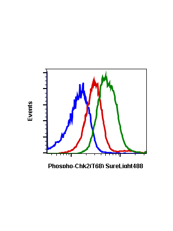
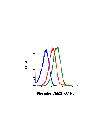
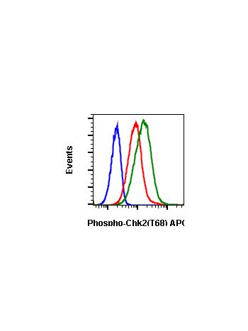
Phospho-Chk2 (Thr68) (D12) rabbit mAb
From
$210.00
In stock
Only %1 left
SKU
2116
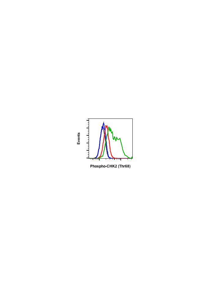
Checkpoint kinase 2 (Chk2) plays a major role in the checkpoint response to DNA damage. Chk2 is initially inactive in its monomeric, unphosphorylated form. Phosphorylation at Thr68 induces homodimerization, initiating autophosphorylation within the kinase loop at Ser516 and phorphorylation events within the auto-inhibitory loop at Thr383 and Thr387. After these phosphorylations, active dimers and monomers can then phosphorylate substrates such as Cdc25C and BRCA1. In humans, Chk2 genetic deletion and missense variants have been found to be associated with increased risk of breast and colon cancer. Constitutively phosphorylated Chk2 at Thr68 has been found in many human cancer cell lines, especially ones with mutations in p53.
| Applications | Flow Cytometry, WB |
|---|---|
| Clone | Chk2T68-D12 |
| Format | Unconjugated |
| Validated Reactivity | Human, Mouse |
| Cross Reactivity | Predicted to work with mouse, rat, and other homologues |
| Detection | Anti-Rabbit IgG |
| Clonality | Monoclonal |
| Immunogen | A synthetic phospho-peptide corresponding to residues surrounding Thr68 of human phospho Chk2 |
| Formulation | 1X PBS, 0.02% NaN3, 50% Glycerol, 0.1% BSA |
| Isotype | Rabbit IgGk |
| Preparation | Protein A+G |
| Recommended Usage | 1µg/mL – 0.001µg/mL. It is recommended that the reagent be titrated for optimal performance for each application. See product image legends for additional information. |
| Storage | -20ºC |
| Pseudonyms | Serine/threonine-protein kinase Chk2, Cds1 homolog, Hucds2, CHEK2, RAD53 |
| Uniprot ID | O96017 |
| References | Ahn J, Urist M, and Prives C. (2004) DNA Repair. 3: 1039-1047. |
Write Your Own Review