Phospho-HS1 (Tyr397) (F12) rabbit mAb
From
$210.00
In stock
Only %1 left
SKU
2396
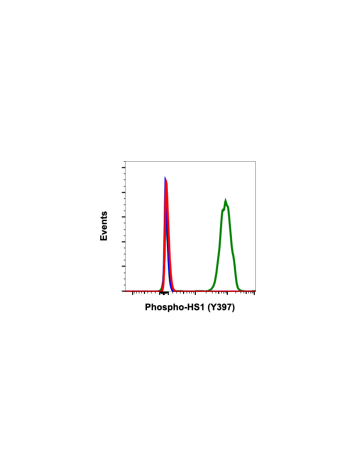
HS1 is expressed in lymphoid and hematopoietic cells, and is heavily post-translationally modified. HS1 deficient mouse models have demonstrated the protein's role in receptor-mediated apoptosis and proliferation. HS1 is phosphorylated at Tyr378 and Tyr397 by the kinase Syk, providing a high-affinity binding site for SH2 domains from the Src family. Following this interaction, HS1 is then phosphorylated at Tyr222 by c-Fgr, Lyn, and Fyn kinases. HS1 plays an important role in T cell signaling, where HS1 phosphorylation recruits and activates Vav1 at the immune synapse. As a homolog of the actin binding protein cortactin, HS1 has been shown to mediate neutrophil chemotaxis through phosphorylation of tyrosines 222, 378, and 397.
| Applications | Flow Cytometry, WB |
|---|---|
| Clone | HS1Y397-F12 |
| Format | Unconjugated |
| Validated Reactivity | Human, Mouse |
| Cross Reactivity | Predicted to work with mouse, rat and other homologues. |
| Detection | Anti-Rabbit IgG |
| Clonality | Monoclonal |
| Immunogen | A synthetic phospho-peptide corresponding to residues surrounding Tyr397 of human phospho HS1 |
| Formulation | 1X PBS, 0.02% NaN3, 50% Glycerol, 0.1% BSA |
| Isotype | Rabbit IgGk |
| Preparation | Protein A+G |
| Recommended Usage | 1µg/mL – 0.001µg/mL. It is recommended that the reagent be titrated for optimal performance for each application. See product image legends for additional information. |
| Storage | -20ºC |
| Pseudonyms | Hematopoietic lineage cell-specific protein, Hematopoietic cell-specific LYN substrate 1, LckBP1, p75, HCLS1 |
| Uniprot ID | P14317 |
| References | Brunati AM, Donella-Deana A, James P, Quadroni M, Contri A, Marin O, and Pinna LA. (1999) Journal of Biological Chemistry. 274:7557-7564. Cavnar PJ, Mogen K, Berthier E, Beebe DJ, and Huttenlocher A. (2012) Journal of Biological Chemistry. 287: 25466-25477. |
Write Your Own Review