SARS-CoV2-2 Iota B.1.526 Variant Recombinant Spike Trimer His Tag
Coronaviruses belong to a large family of protein enveloped positive-strand RNA viruses that are known to cause upper respiratory, gastrointestinal, central nervous system diseases in animals and human (1). One of the main proteins on viral envelop is a heavily glycosylated spike (S) protein (2). Trimeric S protein is involved in host receptor angiotensin-converting enzyme 2 (ACE2) recognition and mediates viral entry into cells. It is the principle antigenic determinant of neutralizing antibodies (3). S protein is recognized by humoral immune response during infection leading to inflammatory response. Homotrimeric S protein is a class I fusion protein that forms large protrusions from the virus surface and undergoes a significant structural rearrangement to fuse the viral membrane with the host-cell membrane once it binds to the host receptor (4).
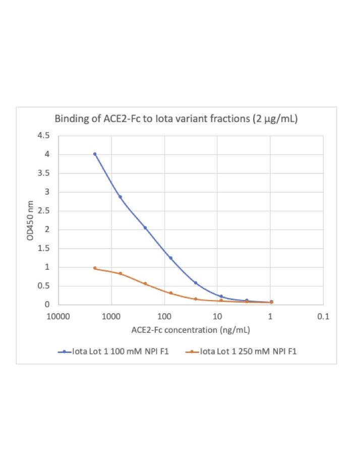
| Applications | ELISA |
|---|---|
| Clone | COV2-Iota |
| Format | His tag |
| Validated Reactivity | Other |
| Cross Reactivity | Predicted to work with mouse, rat and other homologues. |
| Detection | Anti-SARS-CoV-2 mAb |
| Clonality | SARS-CoV-2 |
| Formulation | 1X PBS, 0.025% NaN3 |
| Isotype | SARS-CoV-2 |
| Preparation | His tag purification |
| Recommended Usage | Control antigen for SARS-CoV-2 (COVID-19) diagnostic assays. Contains C-terminal His tag used for purification. |
| Storage | 2-8ºC |
| Pseudonyms | Control antigen for SARS-CoV-2 (COVID-19) diagnostic assays. Contains C-terminal His tag used for purification. |
| Uniprot ID | XXXXXX |
| References | 1. Walls A.C. et al., 2019 Cell, 176: 1026-1039. 2. Tang T. et al., 2020, Antiviral Res., 178:10479. 3. Jiang S et al., 2022, Trends Immunol, 41:355-359. 4. Bosch BJ, et al., 2003, J Virol 77:8801-8811. |