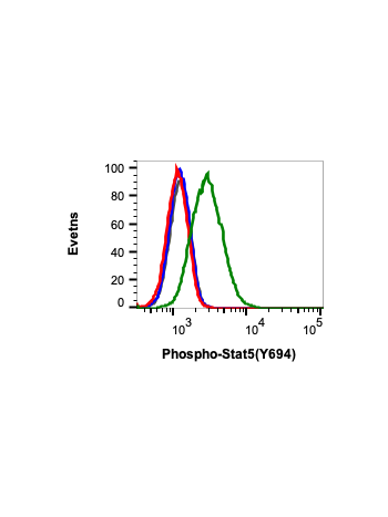
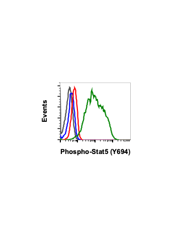
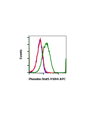
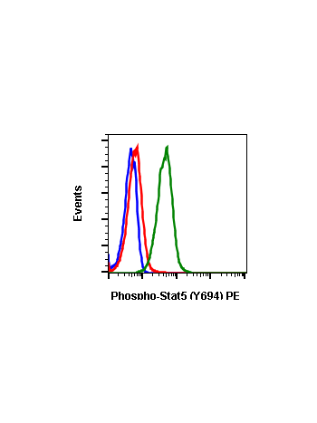
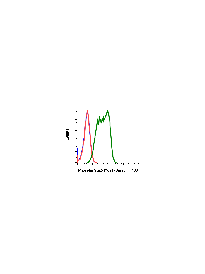
Phospho-Stat5 (Tyr694) (G11) rabbit mAb SureLight 488 conjugate
From
$118.80
In stock
Only %1 left
SKU
2330
Stat5 activation occurs in response to many ligands including prolactin, IL-2, growth hormone, and GM-CSF. Tyr694 phosphorylation is obligatory activation of Stat5 (1,2), and is mediated by Src upon erythropoietin stimulation (3). Phospho Stat5 is constitutively active in some leukemic cell types (4), and phospho Stat5 is found in some endothelial cells when treated with IL-3, suggesting its involvement in cell motility and angiogenesis (5). Stat5 has been shown to be encoded by two separate genes, Stat5a and Stat5b, which share over 90% amino acid sequence identity. In different cell types, Stat5a and Stat5b are independently regulated and activated. For example, interferon treatment predominantly activates Stat5a in U937 cells and Stat5b in HeLa cells (6).
| Applications | Flow Cytometry |
|---|---|
| Clone | Stat5Y694-G11 |
| Format | SureLight 488 |
| Validated Reactivity | Human, Mouse |
| Cross Reactivity | Predicted to work with mouse, rat and other homologues. |
| Clonality | Monoclonal |
| Immunogen | A synthetic phospho-peptide corresponding to residues surrounding Tyr695 of human phospho Stat5 |
| Formulation | 1X PBS, 0.09% NaN3, 0.2% BSA |
| Isotype | Rabbit IgGk |
| Preparation | Protein A+G |
| Recommended Usage | For flow cytometric staining, the suggested use of this reagent is 5 µL per million cells or 5 µL per 100 µL of staining volume. It is recommended that the reagent be titrated for optimal performance for each application. |
| Storage | 2-8ºC |
| Pseudonyms | Signal transducer and activator of transcription 5A, STAT5A, Signal transducer and activator of transcription 5B, STAT5B |
| Uniprot ID | P42229 P51692 |
| References | 1. Gouilleux, F. et al. (1994) EMBO J. 13:4361-4369. 2. Wakao, H. et al. (1994) EMBO J. 13:2182-2191. 3. Okutani, Y. et al. (2001) Oncogene. 20:6643-6650. 4. Demoulin, J.B. et al. (1999) J. Biol. Chem. 274:25855-25861. 5. Dentelli, P. et al. (1999) J. Immunol. 163:2151-2159. 6. Meinke, A. et al. (1996) Mol. Cell. Biol. 16:6937-6944. |
Write Your Own Review